Tyler Morgan: Difference between revisions
mNo edit summary |
|||
| Line 2: | Line 2: | ||
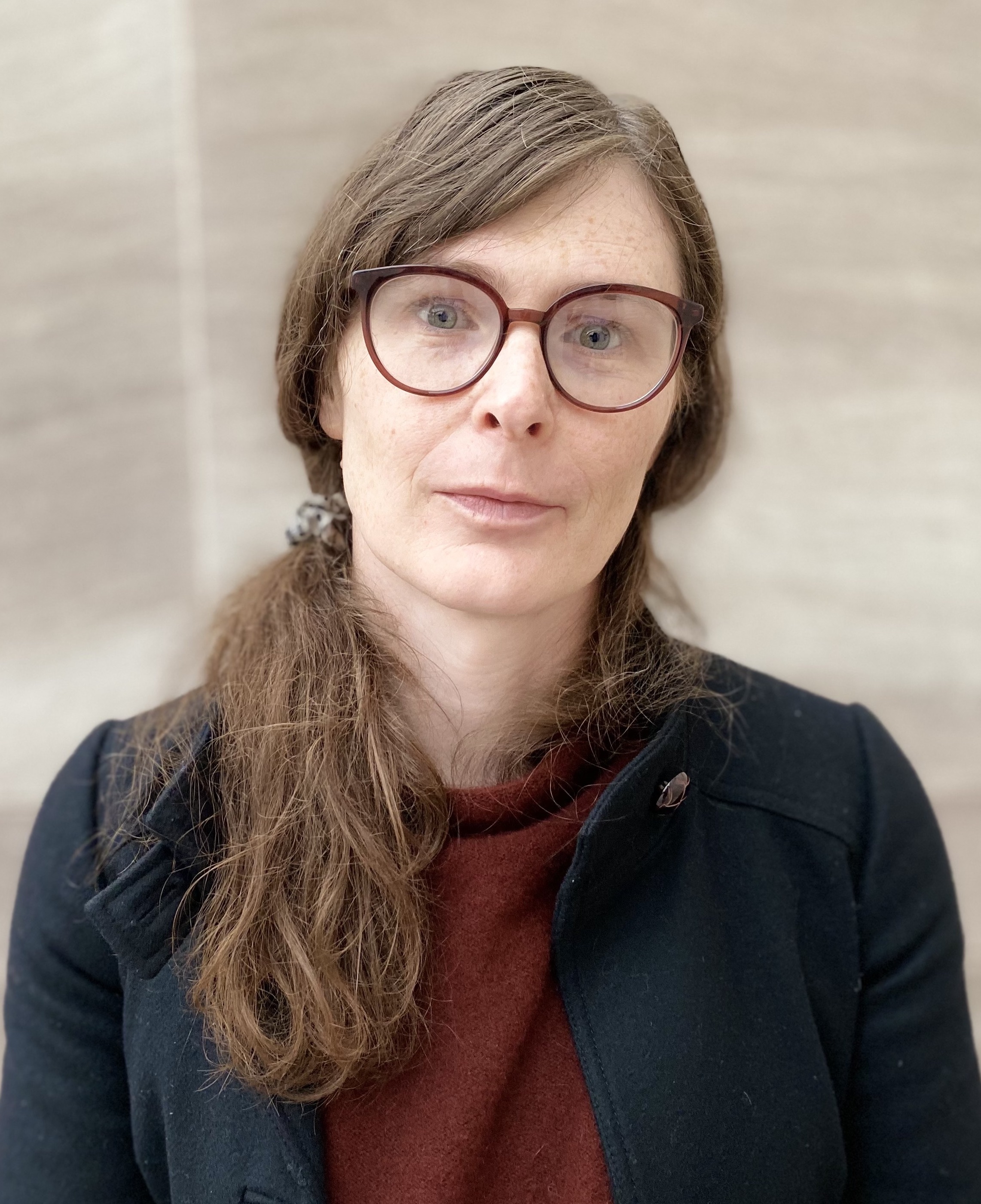
Tyler Morgan has been an FMRIF Staff Scientist since October 2023. She is a neuroscientist and physicist developing high spatial- and temporal-resolution functional imaging methods to measure human brain activity. Her current interests include the development and validation of direct neuronal recordings using MRI (i.e. DIANA signals), as well as development of specialized sequences for hard-to-image regions of the brain. |
Tyler Morgan has been an FMRIF Staff Scientist since October 2023. She is a neuroscientist and physicist developing high spatial- and temporal-resolution functional imaging methods to measure human brain activity. Her current interests include the development and validation of direct neuronal recordings using MRI (i.e. DIANA signals), as well as development of specialized sequences for hard-to-image regions of the brain. |
||
| − | Before joining FMRIF, Tyler was a postdoctoral researcher in the Section on Functional Imaging Methods at NIMH from August 2020. She completed her PhD in Neuroscience & Psychology at the University of Glasgow |
+ | Before joining FMRIF, Tyler was a postdoctoral researcher in the Section on Functional Imaging Methods at NIMH from August 2020. She completed her PhD in Neuroscience & Psychology under Prof. Lars Muckli at the University of Glasgow. Her PhD work focused on predictive and contextual information channels in human cortex and how feedback interacts with feedforward sensory input to create coherent visual perception. |
Revision as of 11:53, 1 March 2024

Tyler Morgan has been an FMRIF Staff Scientist since October 2023. She is a neuroscientist and physicist developing high spatial- and temporal-resolution functional imaging methods to measure human brain activity. Her current interests include the development and validation of direct neuronal recordings using MRI (i.e. DIANA signals), as well as development of specialized sequences for hard-to-image regions of the brain.
Before joining FMRIF, Tyler was a postdoctoral researcher in the Section on Functional Imaging Methods at NIMH from August 2020. She completed her PhD in Neuroscience & Psychology under Prof. Lars Muckli at the University of Glasgow. Her PhD work focused on predictive and contextual information channels in human cortex and how feedback interacts with feedforward sensory input to create coherent visual perception.
Publications
Preprints:
- SI Kronemer, M Holness, AT Morgan, JB Teves, J Gonzalez-Castillo, DA Handwerker, PA Bandettini (2023): Visual imagery vividness correlates with afterimage brightness and sharpness. bioRxiv. doi: 10.1101/2023.12.07.570716.
- ID Driver, RM Sanchez Panchuelo, O Mougin, M Asghar, J Kolasinski, WT Clarke, C Rua, AT Morgan, A Carpenter, K Muir, D Porter, CT Rodgers, S Clare, RG Wise, R Bowtell, ST Francis (2021): Multi-centre, multi-vendor 7 Tesla fMRI reproducibility of hand digit representation in the human somatosensory cortex. bioRxiv. doi: 10.1101/2021.03.25.437006.
- AT Morgan, N Nothnagel, LS Petro, J Goense, L Muckli (2020): High-resolution line-scanning reveals distinct visual response properties across human cortical layers. bioRxiv. doi: 10.1101/2020.06.30.179762.
Published or In Print:
- J Bergmann, LS Petro, C Abbatecola, MS Li, AT Morgan, L Muckli (2024): Cortical depth profiles in primary visual cortex for illusory and imaginary experiences. Nat. Commun. 15, 1002. doi: 0.1038/s41467-024-45065-w
- P Papale, F Wang, AT Morgan, X Chen, A Gilhuis, LS Petro, L Muckli, PR Roelfsema, MW Self (2023): The representation of occluded image regions in area V1 of monkeys and humans. Curr. Biol. 33, 3865-3871. doi: 10.1016/j.cub.2023.08.010.
- M Svanera, AT Morgan, LS Petro, L Muckli (2020): A self-supervised deep neural network for image completion resembles early visual cortex fMRI activity patterns for occluded scenes. J. Vis. 21(7):5, 1–17. doi: 10.1167/jov.21.7.5.
- L Huber, BA Poser, PA Bandettini, K Arora, K Wagstyl, S Cho, J Goense, N Nothnagel, AT Morgan, J van den Hurk, RC Reynolds, DR Glen, R Goebel, OF Gulban (2020): LAYNII: A software suite for layer-fMRI. NeuroImage. doi: 10.1016/j.neuroimage.2021.118091.
- C Rua, WT Clarke, ID Driver, O Mougin, AT Morgan, S Clare, S Francis, K Muir, D Porter, R Wise, A Carpenter, G Williams, JB Rowe, R Bowtell, CT Rodgers (2020): Multi-centre, multi-vendor reproducibility of 7T QSM and R2* in the human brain: results from the UK7T study. Neuroimage. doi: 10.1016/j.neuroimage.2020.117358.
- WT Clarke, O Mougin, ID Driver, C Rua, AT Morgan, M Asghar, S Clare, S Francis, RG Wise, CT Rodgers, A Carpenter, K Muir, R Bowtell (2019): Multi-Site Harmonization of 7 Tesla MRI Neuroimaging Protocols. Neuroimage. doi: 10.1016/j.neuroimage.2019.116335
- AT Morgan, LS Petro, L Muckli (2019): Scene representations conveyed by cortical feedback to early visual cortex can be described by line drawings. J Neurosci. 39(47): 9410-9423. doi: 10.1523/JNEUROSCI.0852-19.2019
- K Ishibashi, CL Robertson, AT Morgan, MA Mandelkern, ED London (2013): The Simplified Reference Tissue Model with 18F-fallypride PET: Choice of Reference Region. Mol Imaging. 12(8). doi: 10.2310/7290.2013.00065.
- M Kohno, DG Ghahremani, AM Morales, CL Robertson, K Ishibashi, AT Morgan, MA Mandelkern, ED London (2013): Risk-Taking Behavior: Dopamine D2/D3 Receptors, Feedback, and Frontolimbic Activity. Cereb Cortex. 25(1):236-245. doi: 10.1093/cercor/bht218.
- DG Ghahremani, B Lee, CL Robertson, G Tabibnia, AT Morgan, N De Shetler, AK Brown, J Monterosso, AR Aron, MA Mandelkern, RA Poldrack, ED London (2012): Striatal dopamine D2/D3 receptors mediate response inhibition and related activity in frontostriatal neural circuitry in humans. J Neurosci. 32(21):7316-24. doi: 10.1523/JNEUROSCI.4284-11.2012.
