Main Page: Difference between revisions
| Line 52: | Line 52: | ||
|[[File:LH Brain Surf.png|alt=LH_Brain_Image|center|frameless|200x200px]] | |[[File:LH Brain Surf.png|alt=LH_Brain_Image|center|frameless|200x200px]] | ||
|[[File:LH Brain Surf.png|alt=LH_Brain_Image|center|frameless|200x200px]] | |[[File:LH Brain Surf.png|alt=LH_Brain_Image|center|frameless|200x200px]] | ||
|[[File: | |[[File:photo_tyler_morgan.png|alt=tyler|center|frameless|200x200px]] | ||
|[[File:LH Brain Surf.png|alt=LH_Brain_Image|center|frameless|200x200px]] | |[[File:LH Brain Surf.png|alt=LH_Brain_Image|center|frameless|200x200px]] | ||
|- | |- | ||
Revision as of 15:20, 3 January 2024
The FMRI Facility (FMRIF)
The functional MRI Facility (FMRIF) is a core resource serving the Intramural Research Program. It was initiated in 1999 primarily by the National Institute of Mental Health (NIMH) and the National Institute for Neurological Disorders and Stroke (NINDS). Its function is to serve as a resource by which all NIH institutes can perform Magnetic Resonance Imaging (MRI) studies that further the understanding of healthy and diseased brain anatomy, function and physiology.
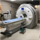
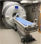
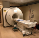
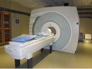
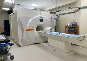
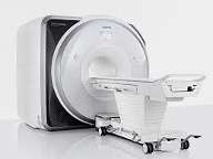
The Facility provides a complete environment for stimulus presentation, monitoring and recording subject behavior and physiology while performing functional MRI (fMRI). At present, the Facility has a total of five scanners for the investigation of humans. These scanners consist of 2 General Electric 3 Tesla MRI scanners, one Siemens 3 Tesla MRI scanner, and 2 Siemens 7 Tesla MRI scanners. More information about each of these scanners and the peripheral user devices equipped can be found here.
| FMRIF
3 Tesla Scanners |
 |
 |
 |
| FMRIF
7 Tesla Scanners |
 |
 |
|
| Collaborative
MRI Scanners |
 |
These currently service the research of about 30 principal investigators in NIMH, NINDS, the National Institute on Alcohol Abuse and Alcoholism (NIAAA), National Institute of Child and Human Development (NICHD), and National Institute of Deafness and Communication Disorders (NIDCD). Since 2000, there have been over 700 papers produced by researchers at the NIH using the core facility scanners and services, with a current paper production rate of about 100 per year.
Other services include scanner operation and instruction, subject interface device development and maintenance, data transfer and storage infrastructure, and multiple web-based services. Our staff scientists also collaborate intensively with the NIH investigators, providing custom capabilities, including pulse sequences, basic processing pipelines, and in-depth advice and consultation. The FMRIF continues to expand as the demand by NIH investigators performing fMRI in their studies continues to grow.
The staff of the FMRIF continues to be committed to maintaining a world-class fMRI scanning environment, providing capability for exploring brain function, physiology, and morphology in healthy volunteers and clinical populations.
Director |
 |
 |
 |
 |
 |
 |
A Brief History of the FMRIF
The FMRIF began in March of 1999 and began operation in September of 2000 when the newly acquired GE 3T scanner first started collecting functional data. In 2002 a second GE 3T started operation. In 2004, a 1.5T scanner devoted to functional MRI of clinical populations was obtained from the NMR center. In 2007, our first 3T scanner was decommissioned to make way for two GE 3T scanners that fit into the same room where one was previously. In 2011, our second 3T scanner was decommissioned to make way for a 7T scanner. Also in 2011, we decommissioned our 1.5T scanner and purchased two state of the art scanners from two different companies: a Siemens Skyra 3T scanner and a GE 750 3T scanner. Starting in late calendar year 2021, FMRIF replaced one of its General Electric 3 Tesla scanners with a Siemens 7T Terra scanner, which was brought online, and opened to users in late 2022. In summary, we now have two GE MR-750 3T scanners, one Siemens Skyra 3T scanner, and two Siemens 7T scanners.
The FMRIF currently has a total of 12 full time employees. This includes five Ph.D. staff scientists, six scanner technologists, and a computer systems analyst, who ensure that the facility provides cutting edge fMRI research capability.
Publications Using Data Collected at the FMRI Facility
-
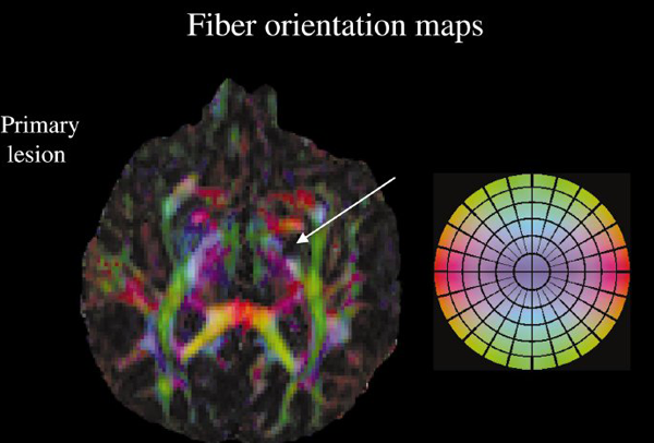
Water diffusion changes in wallerian degeneration and their dependence on white matter architecture. Pierpaoli C, Barnett A, Pajevic S, Chen R, Penix LR, Virta A, Basser P. (2001). Neuroimage. Jun;13(6 Pt 1):1174-85.
-
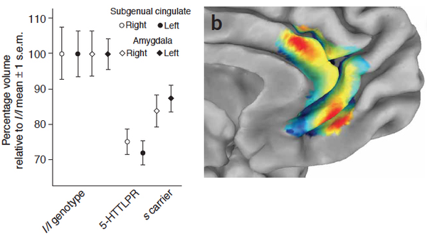
5-HTTLPR polymorphism impacts human cingulate-amygdala interactions: a genetic susceptibility mechanism for depression. In Pezawas L, Meyer-Lindenberg A, Drabant EM, Verchinski BA, Munoz KE, Kolachana BS, Egan MF, Mattay VS, Hariri AR, Weinberger DR. (2005) Nat Neurosci. Jun;8(6):828-34.
-
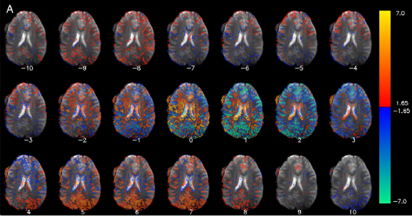
Low-frequency fluctuations in the cardiac rate as a source of variance in the resting-state fMRI BOLD signal. In Pezawas L, Meyer-Lindenberg A, Drabant EM, Verchinski BA, Munoz KE, Kolachana BS, Egan MF, Mattay VS, Hariri AR, Weinberger DR. (2005) Nat Neurosci. Jun;8(6):828-34.
-
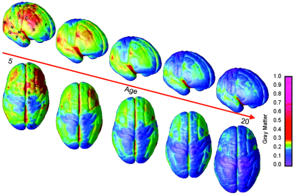
Dynamic mapping of human cortical development during childhood through early adulthood. In Gogtay N, Giedd JN, Lusk L, Hayashi KM, Greenstein D, Vaituzis AC, Nugent TF 3rd, Herman DH, Clasen LS, Toga AW, Rapoport JL, Thompson PM. (2004). Proc Natl Acad Sci U S A. May 25;101(21):8174-9.
-
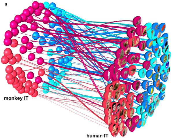
Matching categorical object representations in inferior temporal cortex of man and monkey. In Kriegeskorte N, Mur M, Ruff DA, Kiani R, Bodurka J, Esteky H, Tanaka K, Bandettini PA. (2008) Neuron, Dec 26;60(6):1126-41.

